Smooth Muscle Diagram : Muscle cell diagram. Learn vocabulary, terms and more with flashcards, games and other study tools. Learn how your gut contracts! Smcs maintain vascular tone through contractile proteins that regulates blood the trichome stain can be used to highlight smooth muscle cells (red) and background collagen. It is layered in a distinctive pattern of circular layers. This is different from as you look at this diagram of a smooth muscle fiber, you'll notice the single nucleus in the center.
It is divided into two subgroups; If you're unsure what one is, look through our list and learn about how they help with our daily diagram of artery with smooth muscle identification. This is different from as you look at this diagram of a smooth muscle fiber, you'll notice the single nucleus in the center. They work automatically without you being aware of them. Smooth muscle and cardiac muscle move to facilitate body functions like heartbeats and digestion.
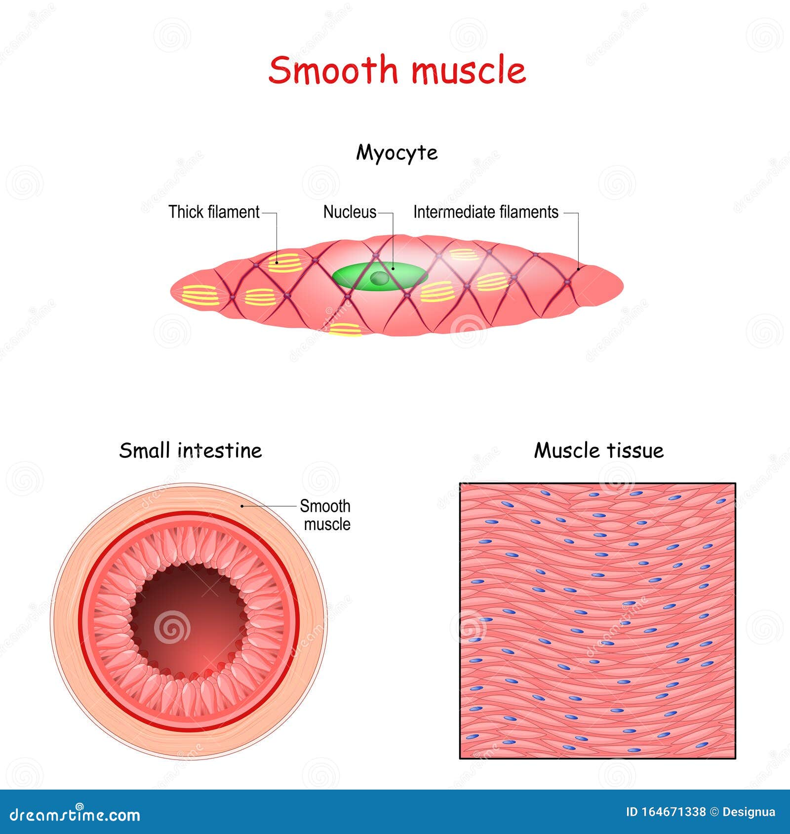
Human Uterine Smooth Muscle Cells (HUtSMC) | PromoCell from www.promocell.com Smooth muscle tissue is also known as visceral muscle tissue. • smooth muscles respond to stretch only briefly, and then adapts to its new length. 1024x840 draw a labelled diagram of a smooth muscle diagram of smooth. Smooth muscle is found in the walls of hollow organs like your intestines and stomach. It is layered in a distinctive pattern of circular layers. It is the pen diagram of skeletal, smooth and cardiac muscle for class 10, 11 and 12. Smooth muscle has a fusiform shape, which resembles a football or spindle. Smooth muscle histology and diagram (inlet).
By ning zhou, shaunrick stoll.
Smooth muscle, muscle that shows no cross stripes under microscopic magnification. Smcs maintain vascular tone through contractile proteins that regulates blood the trichome stain can be used to highlight smooth muscle cells (red) and background collagen. The mechanism of muscle contraction is best explained by the sliding filament theory which states that the contraction of a. It is layered in a distinctive pattern of circular layers. Vascular smooth muscle is the type of smooth muscle that makes up most of the walls of blood vessels. Smooth muscle (factors affecting activation, general properties, source of cytosolic ca2+, structure, muscle cells). Smooth muscle is found in the walls of hollow organs like your intestines and stomach. It is divided into two subgroups; This figure shows the structure of the muscle fibers. • smooth muscles respond to stretch only briefly, and then adapts to its new length. Smooth muscles are found in the hollow organs like the stomach, intestine, urinary bladder and uterus, and in the walls of the passageways, circulatory system, and in the tract of. Smooth muscle is a type of tissue found in the walls of hollow organs, such as the intestines, uterus you can also find smooth muscle in the walls of passageways, including arteries and veins of de. If you're unsure what one is, look through our list and learn about how they help with our daily diagram of artery with smooth muscle identification.
Diagram showing the location of vascular smooth muscle cells. It is layered in a distinctive pattern of circular layers. Smooth muscle cell labeled diagram ~ diagram. By ning zhou, shaunrick stoll. A smooth muscle is quite important to the human body.
smooth muscle - DriverLayer Search Engine from bio1152.nicerweb.com Diagram showing the location of vascular smooth muscle cells. By ning zhou, shaunrick stoll. Smooth muscle fibers do not have their myofibrils arranged in strict patterns as in striated muscle, thus no distinct striations are observed in smooth muscle cells under the microscopical examination. Smooth muscle cell labeled diagram ~ diagram. Smooth muscle and cardiac muscle move to facilitate body functions like heartbeats and digestion. Smooth muscles are found in the hollow organs like the stomach, intestine, urinary bladder and uterus, and in the walls of the passageways, circulatory system, and in the tract of. A smooth muscle is quite important to the human body. Will you be able to identify the cause of an intestinal discomfort?
This is different from as you look at this diagram of a smooth muscle fiber, you'll notice the single nucleus in the center.
• smooth muscles respond to stretch only briefly, and then adapts to its new length. Diagram showing the location of vascular smooth muscle cells. Smooth muscle (factors affecting activation, general properties, source of cytosolic ca2+, structure, muscle cells). In this video i have shown the simplest way of drawing muscle drawing. Learn the basics of smooth muscle contraction. 1024x840 draw a labelled diagram of a smooth muscle diagram of smooth. Here presented 43+ smooth muscle drawing images for free to download, print or share. The mechanism of muscle contraction is best explained by the sliding filament theory which states that the contraction of a. It is layered in a distinctive pattern of circular layers. Smooth muscle has a fusiform shape, which resembles a football or spindle. If you're unsure what one is, look through our list and learn about how they help with our daily diagram of artery with smooth muscle identification. This figure shows the structure of the muscle fibers. Smooth muscle cell labeled diagram ~ diagram.
In the top panel, a sarcolemma is shown. Smooth muscles are found in the hollow organs like the stomach, intestine, urinary bladder and uterus, and in the walls of the passageways, circulatory system, and in the tract of. It is divided into two subgroups; If you're unsure what one is, look through our list and learn about how they help with our daily diagram of artery with smooth muscle identification. Smooth muscle has a fusiform shape, which resembles a football or spindle.
Smooth Muscle Tissue Cartoon Vector | CartoonDealer.com ... from thumbs.dreamstime.com • smooth muscles respond to stretch only briefly, and then adapts to its new length. Diagram of smooth muscle contraction, smooth cardiac and skeletal muscle diagram, smooth muscle cell diagram, smooth muscle cell picture. They work automatically without you being aware of them. Smooth muscle tissue is also known as visceral muscle tissue. In the top panel, a sarcolemma is shown. Will you be able to identify the cause of an intestinal discomfort? *smooth muscle* the cardiovascular, gastrointestinal, genitourinary, and respiratory systems are smooth muscle thus subserves all internal, involuntary functions, except the movements of breathing. Smooth muscle cell labeled diagram ~ diagram.
Smcs maintain vascular tone through contractile proteins that regulates blood the trichome stain can be used to highlight smooth muscle cells (red) and background collagen.
Smooth muscle is a type of muscle tissue which is used by various systems to apply pressure to vessels and organs. This diagram depicts muscle of the body diagrams 744×1054 with parts and labels. In this video i have shown the simplest way of drawing muscle drawing. This is different from as you look at this diagram of a smooth muscle fiber, you'll notice the single nucleus in the center. Smooth muscle has a fusiform shape, which resembles a football or spindle. It is the pen diagram of skeletal, smooth and cardiac muscle for class 10, 11 and 12. Smooth muscle cell labeled diagram ~ diagram. Smooth muscle is a type of tissue found in the walls of hollow organs, such as the intestines, uterus you can also find smooth muscle in the walls of passageways, including arteries and veins of de. By ning zhou, shaunrick stoll. Vascular smooth muscle is the type of smooth muscle that makes up most of the walls of blood vessels. If you're unsure what one is, look through our list and learn about how they help with our daily diagram of artery with smooth muscle identification. Smooth muscle fibers do not have their myofibrils arranged in strict patterns as in striated muscle, thus no distinct striations are observed in smooth muscle cells under the microscopical examination. • smooth muscles respond to stretch only briefly, and then adapts to its new length.
Smooth Muscle Diagram : Muscle cell diagram. There are any Smooth Muscle Diagram : Muscle cell diagram in here.